Chest imaging
Coal mining activities produce coal dust and dust from other minerals, including silica. Coal mine workers who experience repeated exposure to coal mine dust can be at risk of developing coal mine dust lung diseases (CMDLD).
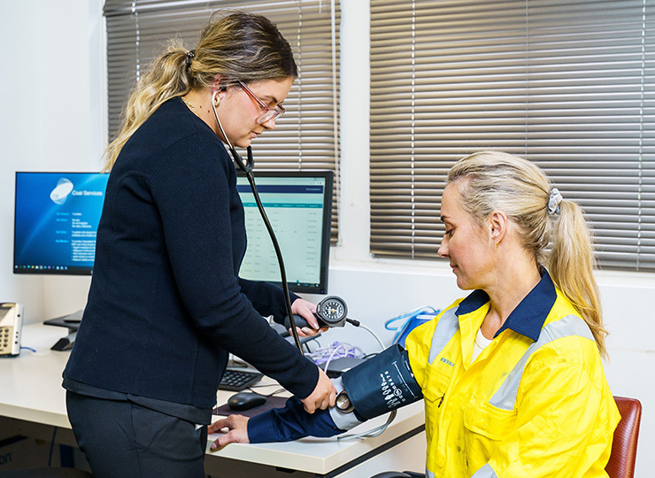
It is important to detect, identify, and diagnose CMDLD as early as possible to support better treatment options and help prevent further lung damage. Early detection relies on several screening tests, including chest imaging.
As part of the Coal Services Health Monitoring Requirements for Coal Mine Workers Order No. 43 (Order 43), CS Health uses two types of chest imaging screening tests: a chest x-ray and/or a high resolution computed tomography (HRCT).
Chest x-ray
A chest x-ray is a painless and non-invasive test that usually takes between 15 and 30 minutes. To capture the images, coal mine workers typically stand in front of an x-ray machine.
In the NSW coal industry, chest x-rays for coal mine workers are reported using the International Labour Organization (ILO) International Classification of Radiographs of Pneumoconioses. This globally-recognised method helps identify and document radiographic changes associated with occupational lung disease.
The chest x-rays are read by radiologists trained in ILO classification, ensuring a consistent and structured approach to identifying abnormalities. This standardised reporting supports early diagnosis, enables approved medical practitioners to monitor changes over time, and ensures timely health assessment reviews when needed. Chest imaging that meets ILO standards forms a critical part of protecting the health of coal mine workers in NSW.
If the chest x-ray shows abnormalities or is inconclusive, an HRCT scan may be recommended for a more detailed view of the lungs and to support further clinical investigations.
HRCT Scan
An HRCT scan is a painless and non-invasive test and takes between 15 and 30 minutes. During the scan, coal mine workers lie on an examination bench that slowly slides into the centre of the HRCT machine while the images are captured.
An HRCT scan is a specialised imaging test that produces detailed images of the lungs and surrounding chest structure, including the lungs and heart. It is used to help detect early signs of occupational lung disease and to assess further lung damage when lung function results are abnormal, or the ILO chest x-ray is inconclusive.
All HRCT scans are classified according to the International Classification of HRCT for Occupational and Environmental Respiratory Diseases (ICOERD). This system ensures consistent interpretation across radiologists, supports accurate documentation of occupational lung disease, and enables better tracking of disease progression. It also helps to inform clinical decisions.
Chest x-ray radiological provider list
Click below to view the chest x-ray radiological provider list.
Share this page